23/09/2026
Epidemiological Evaluation of Gastrointestinal Helminths in 1000 Stray Cats from the Anatolian Side of Istanbul, Türkiye
Abstract
Cats are companion animals that live closely with humans, and parasiticinfections found in cats are important not only for animal health but also forpublic health due to their zoonotic potential. This study aimed to determinethe prevalence of gastrointestinal helminths in fecal samples collectedfrom 1000 stray cats randomly selected on the Anatolian side of Istanbul,a metropolitan city, between June 2022 and February 2025. Fecal samp -les were collected from the ground following natural defecation and exa-mined using native and Fulleborn flotation techniques. Age and sex datawere recorded, and infection rates were statistically compared across thesevariables. Coproscopic analysis revealed that 434 cats (43.4%) were infectedwith at least one helminth. The most commonly detected helminth specieswas Toxocara spp. (25.5%), followed by Toxascaris spp. (4.8%), Dipylidiumspp. (4.2%), Ancylostomatidae eggs (1.2%), Trichuris spp. (0.2%), and Taeniaspp. (0.2%). Among the cats, 36.1% were infected with a single helminthspecies, 6.6% with two species, and 0.7% with three species. No statisticallysignificant difference was found between sexes in terms of parasite preva-lence (p = .769). However, a statistically significant difference was observedamong age groups (p = .001), with a notably higher prevalence in kittensunder six months of age. This study represents one of the most recent andcomprehensive epidemiological investigations with a high sample size ongastrointestinal helminths in stray cats in Istanbul, Türkiye, and the resultsprovide significant contributions to the development of appropriate treat-ment, prevention, and control strategies for cats helminthiasis and raisingawareness of environmental hygiene practices.Keywords: Gastrointestinal helminth, prevalence, stray cat, zoonosis
Introduction
Stray cats are reservoirs for many helminths and may cause infections in humans too, and surveys onhelminths in cats are important not only for their adverse effects on cats but also for human health (Erolet al., 2022;Joachim et al., 2023; Korkmaz et al., 2016). Some of the helminths found in cats are Toxocaraspp., Ancylostomatidae, Dipylidium spp., and Taenia spp. (Joachim et al., 2023; Loftin et al., 2019; Saudaet al., 2019; Sawitri et al., 2024; Villeneuve et al., 2015; Yang & Liang, 2015). One of these, Toxocara cati,is a nematode of cats, and its larvae cause visceral larva migrans in humans (Sauda et al., 2019; Sawitriet al., 2024; Villeneuve et al., 2015; Yang & Liang, 2015). The prevalence of T. cati ranges from 1.7% to 48%worldwide, while in Türkiye it has been reported between 27.8% and 62.5% (Borkataki et al., 2013; Çetin,2023; Ubirajara Filho et al., 2022; Gürler et al., 2015; Henry et al., 2022; Itoh et al., 2012; Iturbe Cossíoet al., 2021; Joachim et al., 2023; Karakuş & Denizhan, 2021; Korkmaz et al., 2016; Little et al., 2015; Loftinet al., 2019; Rabbani et al., 2020; Sauda et al., 2019; Sawitri et al., 2024; Silva et al., 2023; Soe et al., 2023;Soroushianfar et al., 2024; Spada et al., 2013; Villeneuve et al., 2015; Yaman et al., 2006; Yang & Liang,2015). Toxascaris spp., which belongs to the same family (Ascarididae) as T. cati, has been reported tohave a prevalence of 0.2% to 30% worldwide and 1.8% to 9.2% in Türkiye (Çetin, 2023; Gürler et al., 2015;Henry et al., 2022; Itoh et al., 2012; Karakuş & Denizhan, 2021; Rabbani et al., 2020; Soroushianfar et al.,2024; Villeneuve et al., 2015).
Taenia spp. infection in cats is caused by Taenia taeniaformis. Globally, the prevalence of Taenia spp.has been reported to range between 0.3% and 40%, while in Türkiye it ranges between 0.4% and25% (Borkataki et al., 2013; Çetin, 2023; Itoh et al., 2012; Joachim et al., 2023; Little et al., 2015; Loftinet al., 2019; Soe et al., 2023; Spada et al., 2013; Villeneuve et al., 2015; Yaman et al., 2006). Helminthsin Ancylostomatidae are zoonotic hookworms found in cats, especially in warm climates. The infec-tive larvae of these parasites actively penetrate unprotected skin upon contact with contaminated soilor sand, causing cutaneous larva migrans in humans, whereas infection in cats may result in anemia,pale mucous membranes, tachycardia, and lethargy (Bowman et al.,2021). Worldwide prevalence of Ancylostomatidae in cats has beenreported to range between 0.2% and 80%, while in Türkiye it var-ies from 2.7% to 11.94% (Borkataki et al., 2013; Ubirajara Filho et al.,2022; Gürler et al., 2015; Henry et al., 2022; Itoh et al., 2012; Karakuş& Denizhan, 2021; Korkmaz et al., 2016; Little et al., 2015; Loftinet al., 2019; Rabbani et al., 2020; Sauda et al., 2019; Sawitri et al.,2024; Silva et al., 2023; Soe et al., 2023; Spada et al., 2013; Villeneuveet al., 2015; Yang & Liang, 2015). Trichuris spp. infections are gener-ally chronic and more common in adult cats, and prevalences ofinfection are reported between 2.78% and 20.86% worldwide, andbetween 0.46% and 12.5% in Türkiye (Çetin, 2023; Gürler et al., 2015;Mimioğlu, 1951; Soe et al., 2023; Spada et al., 2013; Villeneuve et al.,2015; Yaman et. al., 2006; Yang & Liang, 2015). Dipylidium caninum is a tapeworm transmitted through the ingestion of infected fleas,which act as intermediate hosts. In humans, especially in children,infection can also occur through the accidental ingestion of infectedfleas. Globally, the prevalence in cats ranges from 2.9% to 70%,while in Türkiye it ranges from 5.97% to 12.5% (Borkataki et al., 2013;Çetin, 2023; Henry et al., 2022; Iturbe Cossío et al., 2021; Karakuş &Denizhan, 2021; Little et al., 2015; Loftin et al., 2019; Nourollahi Fardet al., 2024; Rabbani et al., 2020; Sawitri et al., 2024; Silva et al., 2023;Soe et al., 2023; Soroushianfar et al., 2024; Spada et al., 2013; Yamanet al., 2006).The prevalence of gastrointestinal parasites in stray cats variesdepending on several factors, including contact with the externalenvironment, lack of regular antiparasitic treatments, stress levels,age, immune status, and the density of the living environment (Loftinet al., 2019; Sauda et al., 2019). Worldwide studies have reported thatcats, especially those living in shelters or as free-roaming cats, areexposed to many of these risk factors (Baker et al., 2025; Loftin et al.,2019; Villeneuve et al., 2015).Many studies have reported that parasitic infections are common,especially in young cats. The significant decrease in infection prev-alence with advancing age is associated with the development ofimmune responses, acquired resistance, reduced environmentalexposure, and behavioral adaptations (Itoh et al., 2012; Rabbaniet al., 2020; Sauda et al., 2019; Silva et al., 2023; Spada et al., 2013).It is well known that certain helminths, such as Toxocara spp., can betransmitted to kittens via galactogenic or transplacental routes; thisincreases the susceptibility of young individuals to infection (Silvaet al., 2023; Yang & Liang, 2015).
This study not only reveals the prevalence of gastrointestinal hel-minth infections in stray cats but also provides significant data forboth veterinary and public health.
Materials and Methods
Ethics Committee Approval:
All animal procedures were con-ducted in accordance with the EU Directive for animal experiments(European Union, 2010), ARRIVE (Animal Research Reporting of InVivo Experiments) guidelines and national regulation on the protec-tion of experimental animals used for experiments and other scien-tific purposes. Fecal samples were collected from the ground afternatural defecation by the cats during the cleaning of the cage with-out any intervention or handling. The study did not involve any inva-sive procedures on animals; therefore, an ethics committee approvalwas not required.
Sample Collection and Study Design
The study material consisted of fecal samples from 1000 randomlyselected stray cats (499 males, 501 females) that were admitted tothe shelter on the Anatolian side of Istanbul for neutering and vac-cination before being released back to the streets between June2022 and February 2025. The fecal samples were collected fromthe ground after natural defecation by the cats, which were inseparated cages. Approximately 10–15 g of fecal sample from themiddle of the feces on the ground (to minimize the environmentalcontamination risk) was taken from each cat’s feces and stored at+4°C and analyzed for parasitological examination within 24 hours.Data such as age, breed, and sex were recorded from official shelterrecords.The cats included in the study were classified into four groups basedon their ages according to the shelter records. Group A consisted ofcats aged 0–6 months; Group B included cats aged over 6 monthsup to and including 24 months; Group C comprised cats aged over24 months up to and including 84 months (2–7 years); and GroupD included cats aged over 84 months (7 years and older). This agegrouping was performed to evaluate the age-related distribution ofparasitic infection prevalence.
Parasitological Examination:
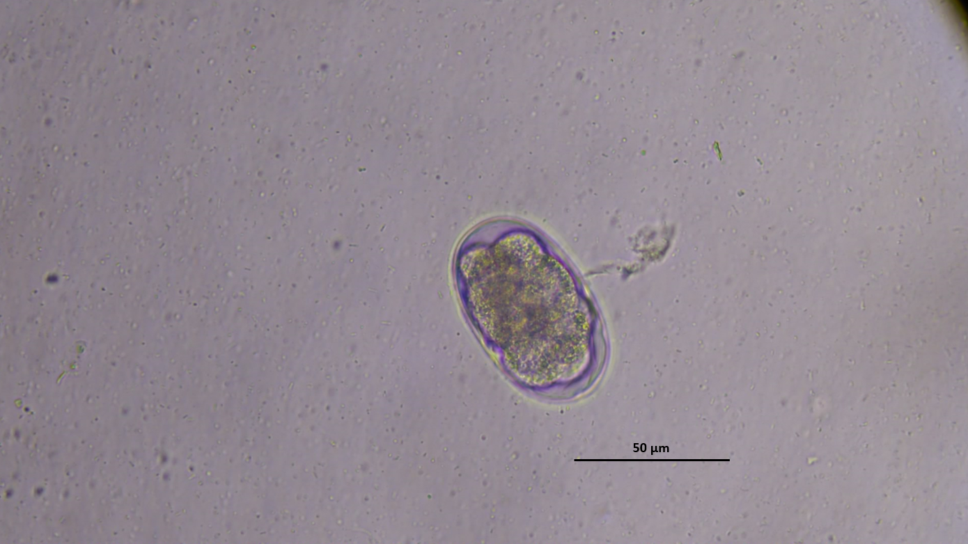
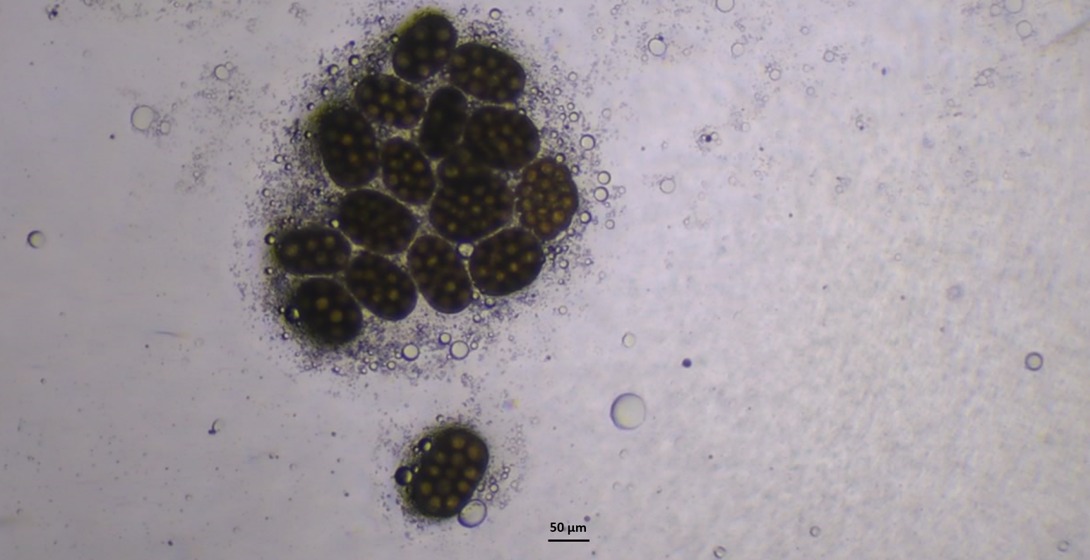
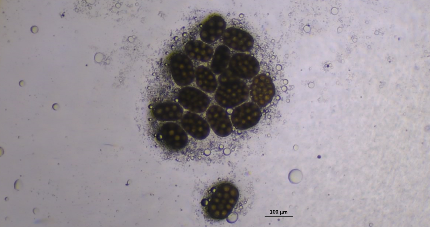
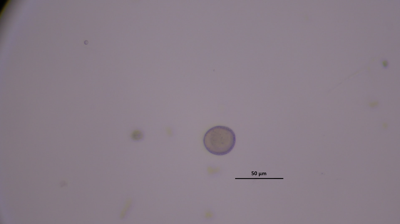
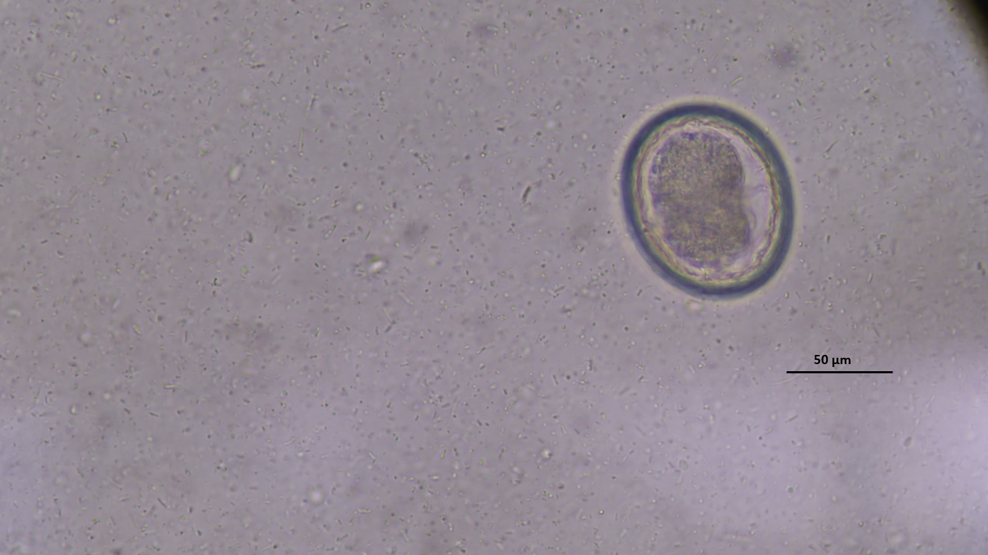
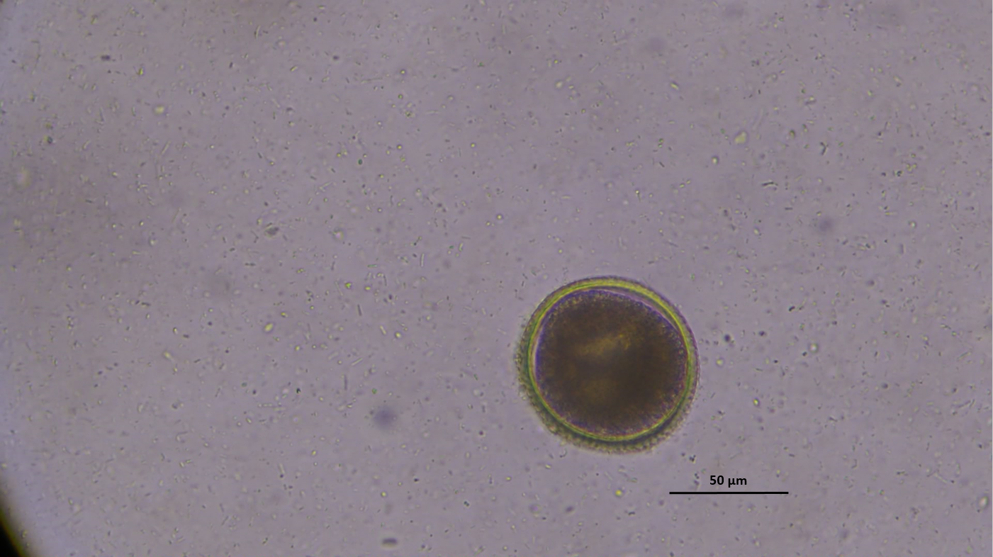
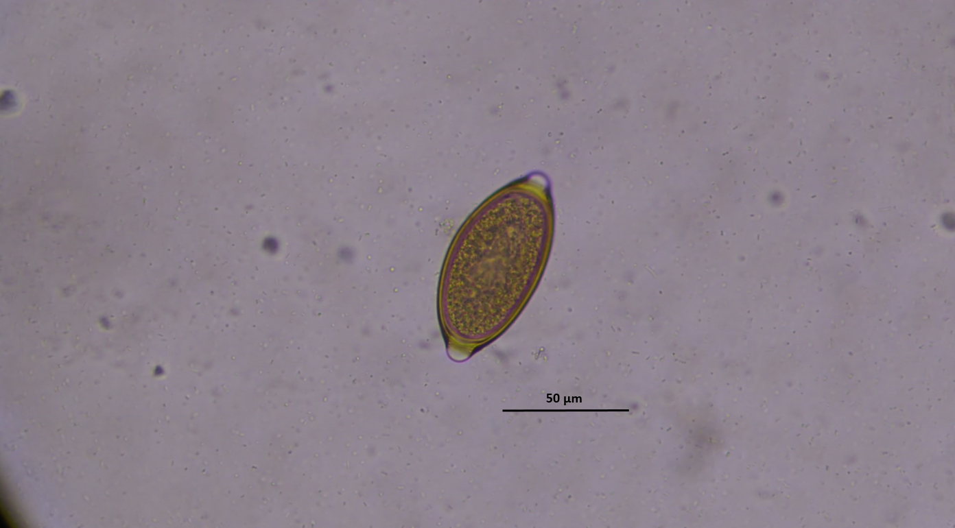
Fecal samples were examined macroscopically for the presence ofadult parasites and cestode proglottids. For native examination, asmall amount of feces was placed on a slide, mixed with a drop oftap water, a cover slip was put on, and the mixture was examinedunder the light microscope. For the flotation technique, approxi-mately 3–5 g of feces was suspended in saturated sodium chloridesolution (specific gravity 1.20), homogenized, and strained througha fine mesh sieve into a clean container. The container was then filledwith the saturated sodium chloride solution until a convex menis-cus was formed, and a coverslip was placed on the solution. After20 minutes, the coverslip was removed and placed on a slide forexamination under 4×, 10×, and 40× objectives of the light micro-scope (Zajac & Conboy, 2012). Helminth eggs were identified basedon their morphological characteristics, including specific structural features, size, and shape as described by Zajac & Conboy (2012).Since there was no objective scale during the microscopic examina-tion, scale bars were added retrospectively to indicate approximateegg size.
Statistical Analysis:
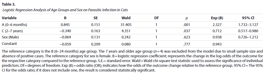
All statistical analyses were performed using IBM SPSS Statistics forWindows, version 26.0 (IBM Corp., Armonk, NY, USA). Descriptivestatistics and the chi-square (χ2) test were used for data analysis. Inthe descriptive analyses, the overall prevalence of parasitic infec-tions and their distribution by sex, age group, and parasitic agentswere expressed as percentages (%) and frequencies (n). To assess theassociation between age, sex, and infection status, a binary logisticregression analysis was performed. Age group was included as acategorical independent variable with three categories, taking the6–24 months age group as the reference. Sex was also included asan independent variable, with female as the reference. The 7 yearsand older age group was excluded from the model due to the smallsample size (n = 4) and the absence of positive cases. The dependentvariable was the presence or absence of parasitic infection. Modelfit was evaluated using the Omnibus Test of Model Coefficients (chi-square test). Statistical significance was set at p < .05.
Results
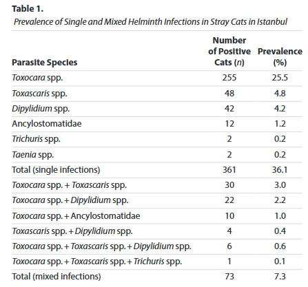
Of the total cat feces samples, 434 (43.4%) were found to be infectedwith at least one parasitic agent belonging to helminths in thisstudy. The most frequently encountered species was Toxocara spp.,found in 25.5% of the cats (Figure 1). Other identified species wereToxascaris spp. (4.8%), D. caninum (4.2%), Ancylostomatidae (1.2%),Trichuris spp. (0.2%), and Taenia spp. (0.2%) (Figures 2–6). Mixedinfections with more than one parasite species were observed in atotal of 73 cats (7.3%). In total, 43.68% (218/499) of male cats and43.11% (216/501) of female cats were found to be infected with par-asitic agents. No significant difference was observed between sexgroups in terms of parasitic infection prevalence (p = .769). The distri-bution and prevalence rates of single and mixed helminth infectionsin stray cats are presented in Table 1.
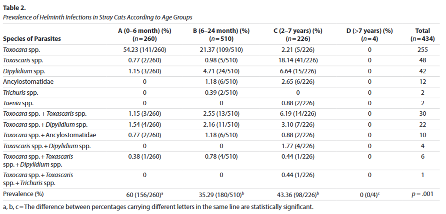
Parasitic infections were notably more prevalent in younger cats,with a significant decline observed as age increased. In group A(n = 260), 60% of the cats tested positive, whereas in group B (n = 510)and group C (n= 226), the prevalence rates were 35.29% and 43.36%,respectively. No parasitic agents were detected in group D. Theprevalence of parasitic infections differed significantly among agegroups (p = .001) (Table 2).
The distribution of various parasitic agents detected in cats withinthe scope of the study was examined according to age groups.Toxocara spp. was identified as the most common parasite, particu-larly more prevalent in younger cats. This species was detected in54.23% (n = 141) of cats in Group A, 21.37% (n = 109) in Group B, andonly 2.21% (n = 5) in Group C. No helminths were found in Group D.Toxascaris spp. showed the highest prevalence in Group C (18.14%,n = 41), whereas D. caninum was most frequently observed in GroupB (4.71%, n = 24). Ancylostomatidae eggs were detected only in Groups B and C, with a prevalence of 1.18% and 2.65% respectively(n = 6). Trichuris spp. was found exclusively in Group B (0.39%, n = 2),and Taenia spp. only in Group C (0.88%, n = 2). Regarding mixed infections, the combination of Toxocara spp. and Toxascaris spp. was most frequently seen in Group C (6.19%, n = 14), followed by GroupB (2.55%, n = 13). The Toxocara spp. + Dipylidium spp. combination was detected similarly in Group A (1.54%), Group B (2.16%), and Group C (3.10%). Toxocara spp. + Ancylostomatidae combinationwas observed in Group A (0.77%), Group B (1.18%), and Group C(0.88%), while the Toxascaris spp. + Dipylidium spp. combinationwas detected only in Group C (1.77%). The triple combination ofToxocara spp. + Toxascaris spp. + Dipylidium spp. was observed in Groups A (0.38%), B (0.78%), and C (0.44%), whereas the combination of Toxocara spp. + Toxascaris spp. + Trichuris spp. was foundonly in Group C (0.44%). No parasitic infections were detected in Group D (the oldest group). These findings indicate that certain par-asites, especially Toxocara spp., are more prevalent in younger agegroups, with a significant decline in infection rates as age increases(Table 2). The binary logistic regression analysis examined the effects of agegroup and sex on the likelihood of parasitic infections in cats. The overall model was statistically significant, indicating that these vari-ables collectively contribute to predicting infection status. Age group was found to be a significant predictor. Specifically, cats aged 0–6months had approximately 2.3 times higher odds of being infectedcompared to cats aged 6–24 months (reference group) (OR = 2.33,95% CI: 1.73–3.13, p = .001). Conversely, cats aged 2–7 years had asignificantly lower likelihood of infection compared to the referencegroup (OR = 0.94, 95% Cl:0.73–1.21, p = .623), including that male andfemale cats had similar odds of infection. These findings suggest thatyounger cats, particularly under 6 months, are at higher risk of para-sitic infection, while older cats between 2 and 7 years have a reduced risk, and gender does not influence the infection rate (Table 3).
Discussion
In the present study, the prevalence of gastrointestinal helminthspecies was 43.4% in the Anatolian side of Istanbul. This prevalenceis quite high and consistent with data reported worldwide and inTürkiye, which generally range between 10.1% and 87.5% (Bakeret al., 2025; Çetin, 2023; Gürler et al., 2015; Henry et al., 2022; Itohet al., 2012; Iturbe Cossío et al., 2021; Joachim et al., 2023; Karakuş &Denizhan, 2021; Korkmaz et al., 2016; Little et al., 2015; Loftin et al.,2019; Rabbani et al., 2020; Sauda et al., 2019; Silva et al., 2023; Soeet al., 2023; Spada et al., 2013; Villeneuve et al., 2015; Yaman et al.,2006, Yang & Liang, 2015).Worldwide studies conducted in countries such as Iran, Brazil,China, Canada, Australia, France, Japan, the United States, Mexico,Myanmar, Indonesia, and Italy have reported Toxocara spp. eggs infeces with prevalence rates ranging from 1.7% to 48% (Borkatakiet al., 2013; Ubirajara Filho et al., 2022; Henry et al., 2022; Itoh et al.,2012; Iturbe Cossío et al., 2021; Joachim et al., 2023; Little et al.,2015; Loftin et al., 2019; Rabbani et al., 2020; Sauda et al., 2019;Sawitri et al., 2024; Silva et al., 2023; Soe et al., 2023; Spada et al.,2013; Villeneuve et al., 2015; Yang & Liang, 2015). Studies con-ducted in Türkiye have reported prevalence rates of Toxocara spp. eggs between 27.8% and 62.5% (Çetin, 2023; Gürler et al., 2015;Karakuş & Denizhan, 2021; Korkmaz et al., 2016; Yaman et al., 2006).In this study, the prevalence of Toxocara spp. was found to be 25.5%,which is consistent with previous findings. Toxocara spp. eggs arehighly resistant to environmental conditions and can survive in soilfor long periods under favorable conditions. Cats tend to bury theirfeces, which may increase the infection risk and make them a res-ervoir for human infections (Joachim et al., 2023; Loftin et al., 2019;Sawitri et al., 2024; Silva et al., 2023; Toparlak et al., 2002; Villeneuveet al., 2015). The prevalence of Toxascaris spp. has been reported torange from 0.2% to 30% worldwide (Henry et al., 2022; Itoh et al.,2012; Rabbani et al., 2020; Soroushianfar et al., 2024; Villeneuveet al., 2015). In studies carried out in Türkiye, the prevalence ofToxascaris spp. has been reported between 1.8% and 9.2% (Çetin,2023; Gürler et al., 2015; Karakuş & Denizhan, 2021). The prevalencerate of 4.8% obtained in this study is within the range reported inprevious studies. The lower prevalence of Toxascaris spp. comparedto Toxocara spp. can be explained by the absence of transplacentaland galactogenic transmission, and the lack of intermediate hosts(Henry et al., 2022; Rabbani et al., 2020; Silva et al., 2023; Villeneuveet al., 2015; Yang & Liang, 2015).Studies conducted globally have reported Ancylostomatidae withprevalence rates ranging from 0.2% to 80% (Borkataki et al., 2013;Ubirajara Filho et al., 2022; Henry et al., 2022; Itoh et al., 2012; Littleet al., 2015; Loftin et al., 2019; Sauda et al., 2019; Sawitri et al., 2024;Silva et al., 2023; Soe et al., 2023; Spada et al., 2013; Rabbani et al.,2020; Villeneuve et al., 2015; Yang & Liang, 2015). In studies con-ducted in Türkiye, prevalence rates have been reported between2.7% to 11.94% (Gürler et al., 2015; Karakuş & Denizhan, 2021;Korkmaz et al., 2016). The prevalence rate of 1.2% found in this studyis within the global range, but notably lower than the rates previouslyreported in Türkiye. The rise in urbanization has led to a reduction insoil-rich areas, resulting in decreased contact between cats and soiland interrupting the transmission cycle of the parasite (Henry et al.,2022; Sawitri et al., 2024; Villeneuve et al., 2015). Worldwide stud-ies have reported the prevalence of D. caninum eggs ranging from2.9% to 70% (Borkataki et al., 2013; Henry et al., 2022; Iturbe Cossíoet al., 2021; Little et al., 2015; Loftin et al., 2019; Nourollahi Fard et al.,2024; Rabbani et al., 2020; Sawitri et al., 2024; Silva et al., 2023; Soeet al., 2023; Soroushianfar et al., 2024; Spada et al., 2013). Studiesconducted in Türkiye have reported Dipylidium spp. prevalence ratesbetween 5.97% and 12.5% (Çetin, 2023; Karakuş & Denizhan, 2021;Yaman et al., 2006). In this study, the prevalence of D. caninum wasfound to be 4.2%.
The prevalence of Taenia spp. has been reported in studies world-wide ranges between 0.3%–40% (Borkataki et al., 2013; Itoh et al.,2012; Joachim et al., 2023; Little et al., 2015; Loftin et al., 2019; Soeet al., 2023; Soroushianfar et al., 2024; Spada et al., 2013; Villeneuveet al., 2015). Studies conducted in Türkiye have reported prevalencerates between 0.4% and 25% (Çetin, 2023; Yaman et al., 2006). Theprevalence rate of 0.2% obtained in this study is lower than thosereported both globally and nationally, indicating a very low preva-lence. Taenia spp. infections occur when cats ingest infected inter-mediate hosts such as rodents, birds, or small mammals duringhunting activities (Loftin et al., 2019; Spada et al., 2013; Villeneuveet al., 2015). In metropolitan cities like Istanbul, stray cats oftenrely on human-provided food sources and commercially preparedpet food. Consequently, their reduced need to hunt decreases therisk of Taenia spp. infection (Loftin et al., 2019; Sawitri et al., 2024).In the present study, Trichuris spp. were identified in 0.2% of theexamined cats. Worldwide studies have documented the preva-lence of Trichuris spp. ranging from 2.78% to 20.86% (Soe et al.,2023; Spada et al., 2013; Villeneuve et al., 2015; Yang & Liang, 2015).The prevalence rate observed in this study is notably lower thanthose reported in the literature. The low prevalence of Trichuris spp.detected in this study may be explained by several factors. In metro-politan areas such as Istanbul, cats often have limited direct contactwith soil, which may reduce the chance of ingesting embryonatedeggs and thus limit the environmental transmission of the parasite(Henry et al., 2022; Spada et al., 2013). The detection of Trichuris spp.eggs is difficult using flotation techniques due to their high specificgravity. However, in cases of severe infection, eggs may rise to thesurface and become detectable by flotation technique (Spada et al.,2013; Villeneuve et al., 2015).
Prevalence rates reported in studies conducted worldwide may varydepending on various factors, such as the living conditions of thecat population (e.g., whether they are household cats or stray cats),environmental factors including climate, temperature, and humidity,the presence or absence of regular antiparasitic treatments, and thediagnostic methods used (Henry et al., 2022; Silva et al., 2023; Soeet al., 2023). Stray cats are more vulnerable than household cats tovarious parasitic infections due to malnutrition, environmental stressfactors, and lack of regular antiparasitic treatments (Spada et al.,2013; Villeneuve et al., 2015).
In this study, the infection rate was 60% in cats aged 0–6 months,35.29% in cats aged 6–24 months, and 43.36% in cats aged 2–7 years,while no parasitic infections were detected in cats older than 7 years.Similar statistically significant differences in the distribution of para-sitic infections among age groups have been reported in previousstudies (p < .05). Particularly, many studies have shown that parasiticinfections are more prevalent in cats under one year of age than inadult cats (Ubirajara Filho et al., 2022; Itoh et al., 2012; Rabbani et al.,2020; Sauda et al., 2019; Silva et al., 2023; Soe et al., 2023; Yang &Liang, 2015). The higher infection rates observed in kittens are likelydue to their still-developing immune systems, which cannot yet offerstrong protection against parasitic agents. In contrast, adult cats maygradually build up a degree of immunity or resistance through con-tinued exposure during their lives (Itoh et al., 2012; Silva et al., 2023;Spada et al., 2013).
Some previous studies have reported a higher prevalence of para-sitic infections in male cats, while others have found it to be morecommon in female cats (Ubirajara Filho et al., 2022; Henry et al., 2022;Silva et al., 2023; Soe et al., 2023). However, these studies did not finda statistically significant difference between sex and parasite preva-lence. Similarly, in this study, sex was not found to have a significanteffect on the prevalence of parasitic infections, and the statisticalanalysis did not reach a significance level (p = .769).
Conclusion
This study revealed the presence of various helminth infections instray cats in Istanbul. These findings will contribute to the develop-ment of appropriate treatment, prevention, and control strategiesfor cats (such as regular antiparasitic treatments and environmental hygiene practices). Since some of these parasites are zoonotic, inves-tigating these parasites is important not only for animal health butalso for public health. However, further studies on different parasitespecies and from various regions need to be investigated.
References
Baker, E., DeBolt, R., Smith, E., Gerhold, R., & Wyrosdick, H. (2025). Fecal preva-lence and diversity of endoparasites in shelter dogs and cats from EastTennessee. Veterinary Parasitology, Regional Studies and Reports, 58,101212.
Borkataki, S., Katoch, R., Goswami, P., Godara, R., Khajuria, J., Yadav, A., &Kaur, R. (2013). Prevalence of parasitic infections of stray cats inJammu, India. Sokoto Journal of Veterinary Sciences, 11(1), 1–6.
Bowman, D. D., Lucio-Forster, A., & Lee, A. C. Y. (2021). Hookworms. In Greene’sinfectious diseases of the dog and cat (5th ed) (pp. 1436–1443). Amster-dam: Elsevier.
Çetin, H. S. (2023). The prevalence of gastrointestinal and pulmonary helmintsin cats on European side of Istanbul and determination of awareness levelof cat owners with regards parasitic diseases [PhD Thesis]. Bursa, Türkiye:University of Uludag.
Erol, U., Altay, K., Şahin, Ö. F., & Urhan, O. F. (2022). Helminth contaminationof commonly consumed raw vegetables in Sivas province in the centralpart of Turkey: first molecular detection of human pathogenic Toxocaracanis eggs in raw vegetables. Acta Veterinaria Eurasia, 49(1), 6-12.
Gürler, A. T., Bölükbaş, C. S., Pekmezci, G. Z., Umur, Ş., & Açıcı, M. (2015). Nema-tode and cestode eggs scattered with cats-dogs feces and significance of public health in Samsun, Turkey. Ankara Universitesi Veteriner FakültesiDergisi, 62(1), 23–26.
Henry, P., Huck-Gendre, C., Franc, M., Williams, T. L., Bouhsira, E., & Lienard, E.(2022). Epidemiological survey on gastrointestinal and pulmonaryparasites in cats around Toulouse (France). Helminthologia, 59(4),385–397.
Itoh, N., Ikegami, H., Takagi, M., Ito, Y., Kanai, K., Chikazawa, S., Hori, Y., Hoshi,F., & Higuchi, S. (2012). Prevalence of intestinal parasites in private-household cats in Japan. Journal of Feline Medicine and Surgery, 14(6),436–439.
Iturbe Cossío, T. L., Montes Luna, A. D., Ruiz Mejia, M., Flores Ortega, A., Here-dia Cárdenas, R., & Romero Núñez, C. (2021). Risk factors associated withcat parasites in a feline medical center. JFMS Open Reports, 7(2),20551169211033183.
Joachim, A., Auersperg, V., Drüe, J., Wiedermann, S., Hinney, B., & Spergser, J.(2023). Parasites and zoonotic bacteria in the feces of cats and dogs fromanimal shelters in Carinthia, Austria. Research in Veterinary Science, 164,105022.
Karakuş, A., & Denizhan, V. (2021). Gastrointestinal parasite infections in catsin Van Province. Van Sağlık Bilimleri Dergisi, 14(2), 191–198.
Korkmaz, U. F., Gökpınar, S., & Yıldız, K. (2016). Prevalence of intestinal para-sites in cats and their importance in terms of public health. Turkiye Par -azitolojii Dergisi, 40(4), 194–198.
Little, S., Adolph, C., Downie, K., Snider, T., & Reichard, M. (2015). High preva-lence of covert infection with gastrointestinal helminths in cats. Journalof the American Animal Hospital Association, 51(6), 359–364.
Loftin, C. M., Donnett, U. B., Schneider, L. G., & Varela-Stokes, A. S. (2019).Prevalence of endoparasites in northern Mississippi shelter cats. Veteri-nary Parasitology, Regional Studies and Reports, 18, 100322.
Mimioğlu, M. (1951). Kedilerimizde müşahede edilen Capillaria aerophila(akciğer kılkurdu) olayları [Cases of Capillaria aerophila (lungworm)observed in cats]. Türk Veteriner Hekimler Derneği Dergisi, 52, 295–299.
Nourollahi Fard, S. R., Akhtardanesh, B., Sadr, S., Khedri, J., Radfar, M. H., &Shadmehr, M. (2024). Gastrointestinal helminths infection of free-roam-ing cats (Felis catus) in Southeast Iran. Veterinary Medicine and Science,10(3), e1422.
Rabbani, I. A. R., Mareta, F. J., Kusnoto, Hastutiek, P., Lastuti, N. D. R., Mufasirin,Suharsono, Sardjana, I. K. W., Sukmanadi, M., & Suwanti, L. T. (2020).Zoonotic and other gastrointestinal parasites in cats in Lumajang, EastJava, Indonesia. Infectious Disease Reports, 12(Suppl. 1), 8747.
Sauda, F., Malandrucco, L., De Liberato, C., & Perrucci, S. (2019). Gastrointesti-nal parasites in shelter cats of central Italy. Veterinary Parasitology,Regional Studies and Reports, 18, 100321.
Sawitri, D. H., Wardhana, A. H., Nefho, F., Purwanto, E. S., Endrawati, D., Nugra-heni, Y. R., Primatika, R. A., Damayanti, N. A., Akbari, R. A., Kusumaning-tyas, E., & Matsubayashi, M. (2024). Prevalence and risk factors associated with zoonotic gastrointestinal helminths transmitted by cats in Jabodetabek, Indonesia. Open Veterinary Journal, 14(10), 2551–2563.
Silva, Y. H. D., Campos, D. R., Lima, G. A. C., Quintal, J. P., Guimarães, B. G., Rêgo,G. M. M. D., Avelar, B. R., Intrieri, J. M., Correia, T. R., & Scott, F. B. (2023).Prevalence of gastrointestinal parasites in domestic cats (Felis catus)diagnosed by different coproparasitological techniques in the munici-pality of Seropédica, Rio de Janeiro. Revista Brasileira de ParasitologiaVeterinaria, 32(3), e006223.
Soe, B. K., Hlaing, K. S., Naing, T. W., Thaw, Z. H., & Myint, A. W. (2023). The firststudy on the prevalence of gastrointestinal parasites in owned and shel-tered cats in Yangon, Myanmar. Veterinary World, 16(2), 414–420.
Soroushianfar, M., Sadr, S., Sazmand, A., Dianaty, S., Khedri, J., Schuster, R. K.,& Borji, H. (2024). Gastrointestinal parasites of cats in the Middle East(2000–2023): a literature review. Parasitology International, 102, 102919.
Spada, E., Proverbio, D., Della Pepa, A., Domenichini, G., Bagnagatti De Giorgi,G., Traldi, G., & Ferro, E. (2013). Prevalence of faecal-borne parasites incolony stray cats in northern Italy. Journal of Feline Medicine and Surgery,15(8), 672–677.
Toparlak, M., Gargılı, A., Tüzer, E., Keleş, V., Ulutaş Esatgil, M., & Çetinkaya, H.(2002). Contamination of children’s playground sandpits with Toxocaraeggs in Istanbul, Turkey. Turkish Journal of Veterinary and Animal Sci-ences, 26(2), 317–320.
Ubirajara Filho, C. R. C. U., Santos, K. K. F., Lima, T. A. R. F., Alves, L. C., Carvalho,G. A., & Ramos, R. A. N. (2022). Gastrointestinal parasites in dogs and catsin line with the one health’ approach. Arquivo Brasileiro de Medicina Vet-erinaria e Zootecnia, 74(1), 43–50.
Villeneuve, A., Polley, L., Jenkins, E., Schurer, J., Gilleard, J., Kutz, S., Conboy, G.,Benoit, D., Seewald, W., & Gagné, F. (2015). Parasite prevalence in fecalsamples from shelter dogs and cats across the Canadian provinces. Para-sites and Vectors, 8, 281.
Yaman, M., Ayaz, E., Gül, A., & Muz, M. N. (2006). Investigation of helminthinfections of cats and dogs in the Hatay Province. Turkish Journal of Para-sitology, 30(3), 200–204.Yang, Y., & Liang, H. (2015). Prevalence and risk factors of intestinal parasitesin cats from China. BioMed Research International, 2015, 967238.
Zajac, A. M., & Conboy, G. A. (2012). Veterinary clinical parasitology (8th ed).Hoboken: Wiley-Blackwell.
Erkınay, S., & Çetinkaya, H. (2026). Epidemiological Evaluation of Gastrointestinal Helminths in 1000 Stray Cats from the Anatolian Side of Istanbul, Türkiye. Acta Veterinaria Eurasia, 52, 1–8. https://doi.org/10.5152/actavet.2026.25156